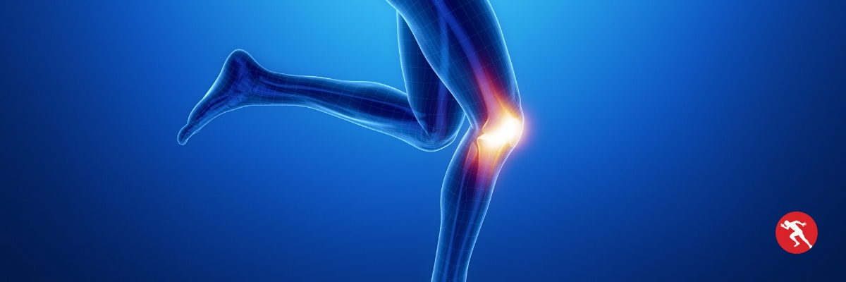
Knee Injuries

Knee injuries are one of the most common injuries we see in people who play sport, run or go to the gym.
There are several different structures in the knee and the best way to describe them would be by location:
- Anterior (front)
- Posterior (back)
- Medial (inner side)
- Lateral (outer side)
- Deep (inside knee)
Physio Performance will identify what is causing your knee injury and how to fix it.
Anterior Knee Injuries
Anterior knee pain can be caused by:
- Patella tendinopathy or tendinitis
- Fat pad impingement
- Patella dislocation or subluxation
- Patellofemoral joint
- Osgood-Schlatter disease
Patellar Tendinopathy
The patella tendon connects the quadriceps muscle to the knee cap (patella).
Sometimes when it gets stressed too much through exercise it can react in two ways:
Tendinitis – the tendon becomes inflamed and painful. The symptoms usually clear up quite quickly with the right treatment and exercises.
Tendinopathy or tendinosis – the tendon thickens over time and becomes painful. There is no inflammation present in this condition. The symptoms usually last for weeks or months.
We see patella injuries mostly in runners, skiers and sports that involve jumping, such as basketball and netball.
This is were it gets the nickname ‘jumper’s knee’.
Fat Pad Impingement or Hoffa's Syndrome
Behind the patellar tendon lies a pad of fat.
It acts to cushion the back of the kneecap against the knee.
It can be very painful.
A physiotherapy assessment will identify what is causing the issue and how to treat it.
Patellofemoral Joint Syndrome or Chondromalacia Patellae
The patella or knee cap has a thin layer of cartilage on the under surface.
This helps the bone to glide in the joint and prevent friction.
Over time this cartilage diminishes, which is part of the normal ageing process.
However, sometimes it can wear away too much due to abnormal stress on the joint from playing sport.
The condition is known as chondromalacia patellae or ‘runner’s knee’.
The condition is best managed through rehabilitation exercises but sometimes athletes require a cortisone injection into the back of the patella for pain relief.
Osgood-Schlatter Disease
A condition that affects active children and adolescents during growth spurts.
The patella tendon attaches to the tibial tuberosity.
The tendon pulls on the attachment on the bone and which causes pain.
Very often a small bump can be seen on the bone due to this repetitive pulling force.
Depending on the severity of pain, children can continue to play sport and manage the condition.
However, some may have to reduce their activity until their growth spurt and pain settles.
Book Expert Knee Injury Assessment And Treatment
Posterior Knee Injuries
Less common is pain at the back of the knee and possible conditions are:
- Baker’s cyst
- Meniscal tear
- Hamstring tendinopathy
- Deep vein thrombosis
Baker's Cyst
A Baker’s cyst is a hernia at the back of the knee that is filled with fluid.
Quite often it looks like swelling or a bulge and it can vary in size.
It can be left alone but if it limits the an athlete’s performance, it can be drained.
However, the swelling can return.
Meniscal Tear
The meniscus is a thick layer of cartilage between the knee joint.
It acts to cushion the joint as we walk, run and jump.
The meniscus can tear if we land and twist on the knee awkwardly.
It can also wear and tear over time due to the ageing process.
When the back of the meniscus (also known as the posterior horn) tears, it can give pain and a feeling of tightness at the back of the knee.
Depending on the size of the injury, the knee joint can lock, click or give way.
In the past most of these knee injuries required arthroscopy (keyhole surgery), but now it is managed through rehabilitation exercises.
Hamstring Tendinopathy
The hamstring tendons pass either side of the back of the knee.
The outer hamstring, known as the biceps femoris is the most common to get injured.
We often see this condition in people training for marathons but it can occur in any sport.
The tendon normally repair themselves after exercise.
However, when the exercise or load on the tendon gets too much, it will become thickened and painful.
It is normally fine at rest but pain occurs during exercise, particularly at the beginning of a session.
People will rest for a long period but when they start exercising again, the symptoms return.
It is important to assess the cause of the problem and then carry out specific rehabilitation exercises to treat it.
Deep Vein Thrombosis (DVT)
Deep vein thrombosis (DVT) is a less common condition seen in active people.
It occurs when a blood clot blocks a vein at the back of the knee or calf.
The area becomes hot, swollen and painful.
If you think you may have a DVT, please seek medical attention as soon as possible.
In athletes, it can occur when the leg is immobilised, in the case of after having recent surgery or the leg in a cast.
Book Expert Knee Injury Assessment And Treatment
Lateral Knee Injuries
The lateral or outer knee has a few different structures that could lead to symptoms.
A detailed physiotherapy assessment is required to diagnose the problem.
The possible injuries are:
- Lateral collateral ligament (LCL) sprain
- Iliotibial band syndrome (ITBS)
- Lateral meniscus
- Lateral hamstring tendinopathy
- Superior tibiofibular joint sprain
Lateral Collateral Ligament (LCL) sprain
The lateral collateral ligament (LCL) provides stability to the outer knee.
It can get stretched by landing awkwardly or from a tackle when playing the sport.
The resulting tear will cause mild, moderate or severe injury.
People sometimes reports a popping sound, instability when trying to walk and swelling.
Iliotibial Band Syndrome
This condition is mostly seen in runners.
The ilitibial band compresses against the outside of the knee during each leg swing when running.
It is caused by abnormal running biomechanics.
The source of the problem can be from weak glutes or pronated feet (low arches).
A cortisone injection is sometimes needed to settle the symptoms and allow the athlete to engage with in a physiotherapy rehabilitation programme to fix the issues.
Meniscal Tear
The meniscus is a thick layer of cartilage between the knee joint.
It acts to cushion the joint as we walk, run and jump.
The meniscus can tear if we land and twist on the knee awkwardly.
It can also wear and tear over time due to the ageing process and develop a cyst.
Depending on the size of the injury, the knee joint can lock, click or give way.
In the past most of these knee injuries required arthroscopy (keyhole surgery), but now it is managed through rehabilitation exercises.
Lateral Hamstring Tendinopathy
The outer hamstring, known as the biceps femoris can developed symptoms on the outer knee.
We often see this condition in people training for marathons but it can occur in any sport.
The tendons normally repair themselves after exercise.
However, when the exercise or load on the tendon gets too much, it will become thickened and painful.
It is normally fine at rest but pain occurs during exercise, particularly at the beginning of a session.
People will rest for a few weeks but when they start exercising again, the symptoms return.
It is important to assess the cause of the problem and then carry out specific rehabilitation exercises to treat it.
Superior Tibiofibular Joint
The fibula is a long bone on the outer part of your lower leg.
It extends from the ankle right up to the outer knee.
The head of the bone feels like a hard marble on the outside of the knee.
This joint has ligaments and cartilage.
It can become sprained due to direct contact, in a tackle for example.
It can also be irritated by abnormal running biomechanics.
The common peroneal nerve injury wraps around the head of the fibula.
It can also be injured by direct contact, resulting in pain, pins and needles or numbness in the lower leg.
Book Expert Knee Injury Assessment And Treatment
Medial Knee Injuries
The medial or inner knee has a few different structures.
These medial knee injuries would be more common than lateral knee injuries.
Possible injuries are:
- Patellofemoral joint syndrome
- Medial collateral ligament (MCL) sprain
- Medial meniscus injury
- Osteoathritis
- Pes anserius bursitis
Patellofemoral Joint Syndrome or Chondromalacia Patellae
The patella or knee cap has a thin layer of cartilage on the under surface.
This helps the bone to glide in the joint and prevent friction.
Over time this cartilage diminishes, which is part of the normal ageing process.
However, sometimes it can wear away too much due to abnormal stress on the joint from playing sport.
The condition is known as chondromalacia patellae or ‘runner’s knee’.
The condition is best managed through rehabilitation exercises but sometimes athletes require a cortisone injection into the back of the patella for pain relief.
Medial Meniscal Tear
The medial meniscus is a thick layer of cartilage between the knee joint.
It acts to cushion the joint as we walk, run and jump.
The meniscus can tear if we land and twist on the knee awkwardly.
It attaches onto to medial collateral ligament (MCL) so it is possible to injure both the meniscus and MCL at the same time.
It can also wear and tear over time due to the ageing process and develop a cyst.
Depending on the size of the injury, the knee joint can lock, click or give way.
In the past most of these injuries required arthroscopy (keyhole surgery), but now it is managed through rehabilitation exercises.
Knee Osteoarthritis
Osteoarthritis is a normal part of the ageing process.
It is like our hair turning grey or developing wrinkles on our skin.
The knee has a thick meniscus between the bones and a thin layer of cartilage (articular cartilage) covering the bone to protect the joint.
Over time the meniscus and articular cartilage can become thinner, resulting in more pressure on the bone ends.
There is more surface area and pressure on the inner part of the knee in comparison to the outer.
This makes the medial side of the knee more prone to osteoarthritis, which can cause pain and swelling in that area.
This condition is managed through pain relieving treatment and strength exercises.
If the osteoarthritis is severe and debilitating, a knee replacement is offered to replace the bone surfaces.
Pes Anserius Bursitis
The medial side of the knee has three muscle attachments (sartorius, gracilis and semomembranosus).
Between the tendons and the bones lies a bubble of fluid called a pes anserius bursa.
This helps to prevent friction.
It can become inflammed and painful in sports such as swimming (especially breaststroke), cyclists and runners.
Book Expert Knee Injury Assessment And Treatment
Deep Knee Injuries
Injuries deep inside the knee are known as ‘intra-articular’ injuries.
They normally present with widespread or general pain and swelling.
However, structures such as the anterior cruciate ligament or posterior meniscus can result in posterior or behind the knee pain.
Possible injuries include:
- Anterior cruciate ligament tear
- Posterior cruciate ligament tear
- Meniscal tear or degeneration
- Osteochondral defect
Anterior Cruciate Ligament (ACL) Tear
The anterior cruciate ligament (ACL) provides stability to the knee joint.
Injuries to the ligament are often seen in sports that involve pivoting or twisting on the knee i.e. football, basketball, American football, Australian Rules football, gymnastics and skiing.
However, it can also occur through direct contact. For example, when an athlete’s foot in planted on the ground and an opposing player collides with the knee.
The injury can occur in combination with others, such as a meniscal and medial collateral ligament (MCL) tear.
Unfortunately, a complete tear to the ACL requires surgery and the recovery time is at least 9 months, before returning to sport.
A portion of the hamstrings or patella tendon is used as a graft to replace the ACL.
Posterior Cruciate Ligament (PCL) Tear
The posterior cruciate ligament (PCL) sits close to the ACL deep inside the knee but it is less commonly injured than the ACL.
It also can be ruptured by twisting injuries of the knee or through direct contact, such as a tackle.
Not all PCL injuries require surgery.
A physiotherapy programme focusing on strengthening the knee and improving movement patterns such be enough.
However, if there is significant instability or other associated injuries that could limit recovery, then surgery would be indicated.
Meniscal Tear
We have already discussed meniscal injuries in the medial and lateral injury section.
The pain is commonly either on the outside or inside of the knee but it can also present as a general ache, deep inside the knee.
The menisci are two C-shaped thick layers of cartilage between the knee joint.
They act to cushion the joint as we walk, run and jump.
The menisci can tear if we land and twist on the knee awkwardly.
It can also wear and tear over time due to the ageing process and develop a cyst.
Depending on the size of the injury, the knee joint can lock, click or give way.
In the past, meniscal injuries required arthroscopy (keyhole surgery) to fix them.
Nowadays they are managed through physiotherapy and rehabilitation exercises.
Surgery is required if rehabilitation has been unsuccessful or the tear is large, leading to the joint locking.
Osteochondral Defect
In addition to the menisci providing cushioning and support to the knee joint, the bones are also covered in a thin layer of cartilage called articular cartilage.
Damage to the articular cartilage often occurs in combination with other knee injuries such as an ACL tear.
The force from the injury breaks off a piece of the cartilage, resulting in a ‘loose body’ floating around the knee.
This is known as an osteochondral defect.
Symptoms include knee pain, swelling and locking.
Depending on the degree of the injury, these can be managed conservatively or may require surgery depending the severity.
Surgery involves removing the loose body and repairing the area of the bone where of piece of cartilage broke away from.
At our Newforge Clinic we provide a range of treatments including physiotherapy, sports massage, acupuncture and dry needling. We treat many common conditions including lower back pain, shoulder injuries, neck injuries and shin splints.
